Elizabeth Jekot, MD, has a family history of breast cancer.
"That afforded me a new sense of empathy for my patients and their family members," says Dr. Jekot, who serves as the medical director of the Elizabeth Jekot, MD Breast Imaging Center at Baylor Scott and White Medical Center — Plano.
Yet, even with the advancements in detection imaging, the misconceptions around mammography may keep some women from finding the answers they need — answers that could save their lives, especially when found early.

The most common misconceptions that Dr. Jekot encounters stem from fear. It's fear on many levels. First, there's the fear that the patient has of finding something or of entertaining a false sense of security if the mammogram somehow doesn't "catch" their cancer.
What Dr. Jekot tries to explain to such patients is this: the mammogram is an excellent tool — especially with the ongoing development of 3D imaging — but it is not perfect.
"When we look at a mammogram, we only get two colors that show up: gray and white," Dr. Jekot explains. "The gray is fat, and white is normal tissue. It's the ratio of gray fat to white tissue that determines your density. Like anything in life, it's like a spectrum. There is also a population of patients that have what we call a dense breast tissue; for those patients, the mammogram is not as sensitive as for other patients."
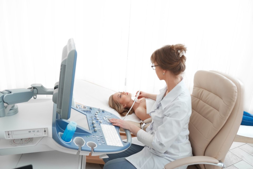
But there is some hope for those patients, Dr. Jekot explains. In Texas, Henda's Law offers additional screening protocols that are more sensitive for patients with more dense breast tissue. Some of these protocols include offering magnetic resonance imaging (MRI) or a bilateral breast sonogram.
The next fear Dr. Jekot sees in patients is the fear of discomfort or pain, typically due to disturbing stories patients might have heard or read about. For some, it can be uncomfortable, but it shouldn't hurt.
Dr. Jekot gives a checklist for women who are going in for a mammogram, whether it's their first or tenth time, to help assuage their fear and provide the best imaging experience possible.
- Have your annual screening once you hit the age of 40. This applies to average patients without overriding factors such as family history.
- Faithfully practice your monthly self-exam, keeping time in mind. Perform this exam preferably 5 to 7 days before your period starts. This timeline goes for scheduling your mammogram too! It doesn't matter if you're post-menopausal though. Timing your mammogram appointment can determine how uncomfortable you experience may be, too.
- Get a regular doctor's clinical exam. This goes hand-in-hand with your self-exam.
- Wear a comfortable two-piece outfit when you get imaged. No dresses, just so that you don't feel as awkward or vulnerable during the process.
- Remember, the compression only lasts a few seconds. It really can be a tolerable exam with minimal discomfort. The technologist is on your side to get the best possible imaging.
- Relax, relax, relax! Tense patients tend to be far more uncomfortable during the process
For more information or to schedule a mammogram with Baylor Scott & White Health, please call 214-865-4000, or go online to BSWHealth.com/BreastImaging.


